What is Mohs Surgery?
In the early 1940s, Frederick Mohs, professor of surgery at the University of Wisconsin, developed a form of treatment for skin cancers he called chemosurgery.
“Chemosurgery” is derived from the words “chemical” and “surgery.” The addition of “Mohs” honors the doctor who developed the technique. It is a highly specialized form of treatment for the total removal of skin cancers. It is performed by a team of medical personnel that includes physicians, technicians, and nurses or medical assistants.
Dr. Mehrany, who is heading the team, is board certified in Micrographic Dermatologic Surgery after having had subspecialty fellowship surgical training in Mohs micrographic & reconstructive surgery techniques. A technician, whom you may not even meet, performs the important task of preparing the tissue slides, which are examined under a microscope by Dr. Mehrany.
The word “chemosurgery,” when used today, is really a misnomer. When Dr. Mohs initially introduced the procedures, he applied a chemical (zinc chloride) to the tumor and surrounding skin, which fixed the tissue prior to its removal. Since 1974, the procedure has been refined and improved upon, so the vast majority of cases are done using fresh tissue (omitting the chemical paste).
Although the official name for the procedure is Mohs micrographic surgery, we prefer the shortened version of Mohs Surgery. The surgery is performed as follows: the skin suspicious for cancer is treated with a local anesthetic, so there is no feeling of pain in the area. To remove most of the visible skin cancer, the tumor is scraped using a sharp instrument called a curette. A thin piece of tissue is then removed surgically around the scraped skin and carefully divided into pieces that will fit on a microscope slide; the edges are marked with colored dyes; a precise map or diagram of the tissue removed is made; and the tissue is frozen by the technician. Thin slices can then be made from the frozen tissue and examined by the doctor under the microscope.
Most bleeding is controlled using light cautery, although occasionally, a small blood vessel is encountered, which must be tied off with suture. A pressure dressing is then applied, and the patient is asked to wait while the slides are being processed. Dr. Mehrany will then examine the slides under the microscope to determine if any cancer is still present and subsequently annotate his map of the cancer location accordingly. If cancer cells remain, they can then be located at the surgical site on the patient by referring to the map. Another layer of tissue is then removed, and the procedure is repeated until Dr. Mehrany is satisfied that the entire base and sides of the wound have no cancer cells remaining. As well as ensuring total removal of cancer, this process preserves as much normal, healthy surrounding skin as possible.
The removal and processing of each layer of tissue take approximately 1-3 hours. Only 20 to 30 minutes of that is spent in the actual surgical procedure. The remaining time is required for slide preparation and interpretation. It usually takes the removal of two or three layers of tissue (also called stages) to complete the surgery. Therefore, by beginning early in the morning, Mohs surgery is typically finished in one day. At the end of Mohs surgery, you will be left with a surgical wound free of tumor which is then reconstructed that same day. Several options for reconstruction may be discussed with you in order to weigh your preference of what will provide the best possible cosmetic outcome versus what will provide the easiest and simplest recovery.
For more information click the Link Below
The Reconstruction Possibilities Include:
Healing by spontaneous granulation involves letting the wound heal by itself. This offers a good chance to observe the wound as it heals after the removal of a difficult tumor. Experience has taught us that there are certain areas of the body where nature will heal a wound as nicely as any further surgical procedure. There are also times when a wound will be left to heal, knowing that if the resultant scar is unacceptable, some form of reconstructive surgery can be performed at a later date.
Closing the wound with stitches is often performed on a small lesion. This involves some adjustment of the wound and sewing the skin edges together. This procedure speeds healing and can offer a good cosmetic result. For example, the scar can be hidden in a wrinkle line.
Skin grafts are another reconstruction option which involve covering a surgery site. There are two types of skin grafts. The first is called a split-thickness graft. This is a thin shave of skin, usually taken from the thigh, which is used to cover a surgical wound. This can be either permanent coverage or temporary coverage before another cosmetic procedure is done at a later date. The second graft type is the full-thickness graft. This graft requires a thicker layer of skin to achieve proper results. In this instance, skin is usually removed from around the ear or collarbone (the donor site) and stitched to cover a wound. The donor site is then sutured together or left alone to be allowed to heal on its own depending what will provide a superior cosmetic outcome.
Skin flaps involve the movement of adjacent, healthy tissue to cover a surgical site. Where practical, they are chosen because of the excellent cosmetic match of nearby skin. If your Mohs Surgery is extensive or performed where a functional impairment results, we may recommend you visit one of several consultant physicians. If you have been sent to us by a physician skilled in skin closures (for example, a plastic or reconstructive surgeon), he or she may take care of you after your cancer has been removed.
In summary, by microscopically pinpointing affected skin cancer areas and removing these tissues, Dr. Mehrany can successfully remove your skin cancer. Because normal tissue is preserved to the greatest extent possible, Dr. Mehrany is able to offer you the possibility of an optimal cosmetic result. Although the fullest attempt will be made to minimize the scar, there is always a scar with surgery. There is no such thing as scar-less surgery.
Advantages of Mohs Surgery
Mohs Micrographic Surgery is an advanced treatment procedure for skin cancer, which offers the highest potential for recovery – even if the skin cancer has previously been treated. This procedure is a state-of-the-art treatment in which the physician serves as the Mohs surgeon, pathologist, and reconstructive surgeon. The technique allows for the highest possible cure rates while conserving the most tissue to ensure the best possible cosmetic outcome. This procedure allows dermatologists trained in Mohs Surgery to see beyond the visible disease and to precisely identify and remove the entire tumor, leaving healthy tissue unharmed.
Mohs surgery is most often used in treating two of the most common forms of skin cancer: basal cell carcinoma and squamous cell carcinoma. The cure rates for Mohs Micrographic Surgery are the highest of all treatments for skin cancer -- up to 99 percent, even if other forms of treatment have failed. This procedure is the most exact and precise method of cancer removal, minimizing the chances of cancer re-growth and decreasing the potential for scarring or disfigurement.
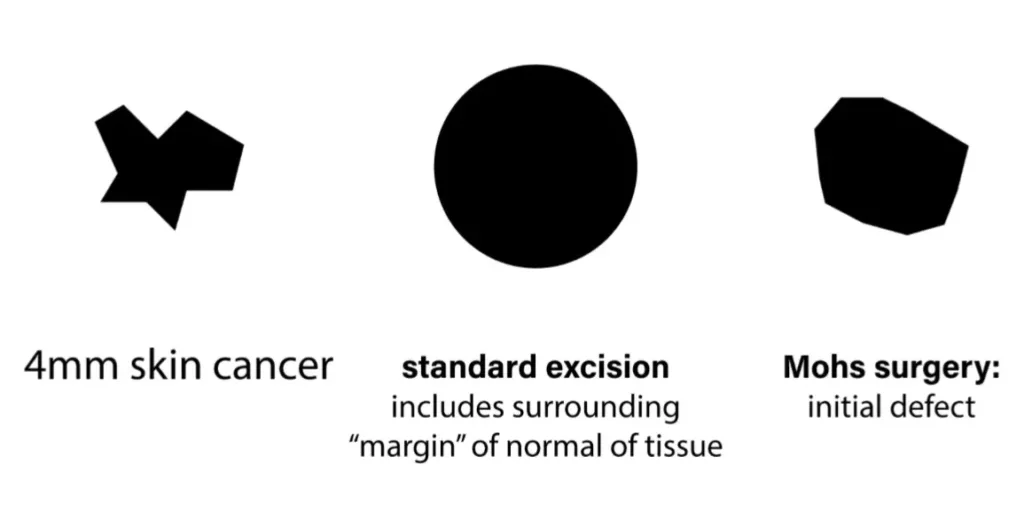
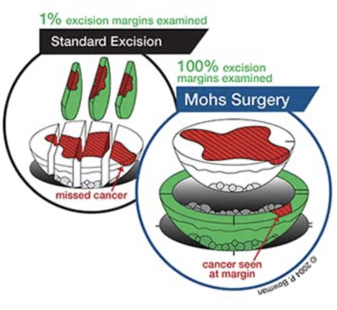
Mohs surgery is different from other skin cancer treatments because only cancerous tissue is removed. The Mohs surgeon does not take an additional “margin” of normal tissue in order to make sure “we get it all”. Only in Mohs surgery is 100% of the complete and entirety of the excised tissue edges microscopically examined. This is in stark contrast to standard non-Mohs excisions where less than 1% of the excised tissue edges are microscopically examined. As exhibited in the diagram the manner in which the tissue is removed, processed and examined is completely different in Mohs vs. standard excision.


Common Indications for Mohs Surgery Include:
- The cancer is in an area where it is important to preserve healthy tissue for a maximal functional and cosmetic outcome, such as head & neck, eyelids, nose, ears, lips, cheeks, chin, forehead, hands, feet or genitalia.
- The cancer is not clearly defined at the edges
- The cancer is large
- The cancer was treated previously and recurred
- The cancer displays aggressive pathology
- The cancer is in an area where scar tissue is present
- The cancer is growing rapidly or uncontrollably
- Mohs surgery is cost-effective. This technique was found to be similar in cost to other procedures, such as electrodesiccation and curettage, cryosurgery, excision, or radiation therapy. Mohs surgery preserves the maximum amount of normal skin, leading to simpler repairs and fewer complicated reconstructive procedures. With its high cure rate, Mohs surgery minimizes the risk of recurrence and eliminates the costs of larger, more serious surgery for recurrent cancers.
